Quick takeaways
- New imaging tools are making scans sharper, faster, and more information-rich.
- AI can help process images, but the scan still has to answer a real clinical question.
- Better imaging is useful when it changes diagnosis, treatment, or follow-up.
Medical imaging comes with an alphabet soup: MRI, CT, PET, SPECT and DEXA. I used to think progress meant inventing a brand-new scanner. A lot of the interesting work now is less flashy. It makes the machines hospitals already use quicker, easier to move, or better at pulling a useful image from imperfect data.
That is where AI and new detector hardware start to overlap. A scan is not just a picture anymore. Software can reconstruct it, clean it up, measure things in it and sometimes guide the scan as it happens. The risk is assuming that a sharper or faster image automatically leads to better care.
Photon-counting CT is the cleanest hardware story
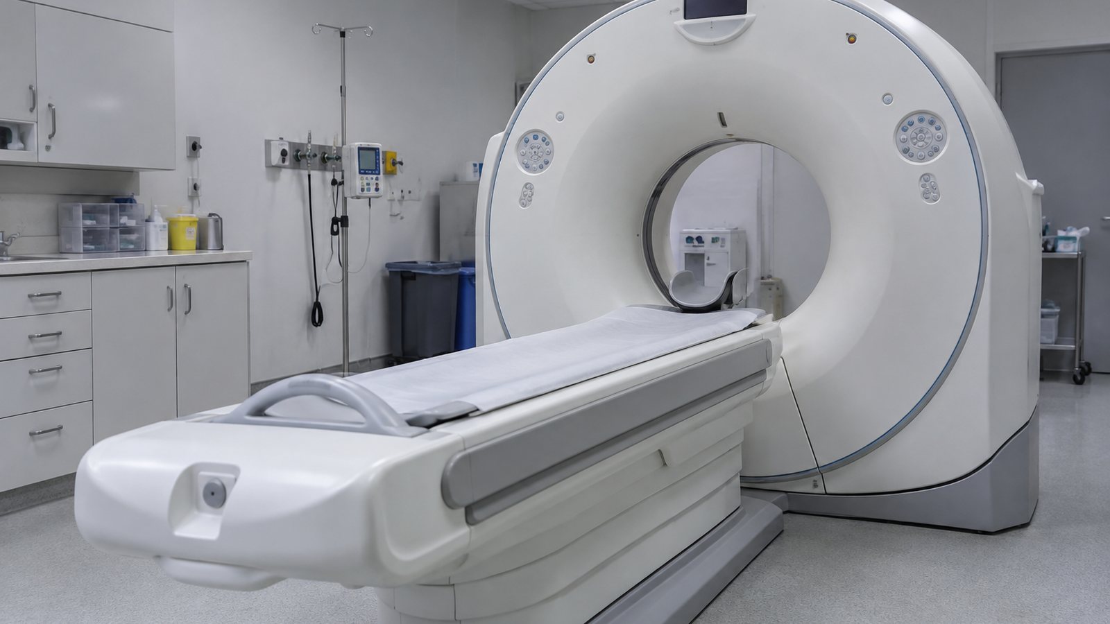
A standard detector adds up the energy that reaches it. A photon-counting detector records individual X-ray photons and their energy. In the review by Willemink and colleagues, that design improved spatial resolution and helped separate materials in the image (Willemink et al.). It may also reduce the radiation or contrast dose needed for some scans.
The clinical version became real when Siemens Healthineers received FDA clearance for the NAEOTOM Alpha in 2021, described as the first photon-counting CT scanner cleared for clinical use in the United States (Siemens Healthineers). The technology is now clinical equipment, though most hospitals do not have it.
If CT can show smaller structures with less radiation or less contrast dye, it may help patients who need repeat scans, people with kidney concerns and patients whose care depends on tiny anatomical changes. The useful version gives the radiologist more information with less burden on the patient.
MRI is getting faster through reconstruction
MRI has a different problem. Although it does not use ionizing radiation, it can be slow, loud and hard for patients who are claustrophobic or unable to hold still. Traditionally, faster MRI meant collecting less data and accepting worse image quality. AI reconstruction tries to change that tradeoff.
Hammernik et al. published a variational network approach for accelerated MRI reconstruction in Magnetic Resonance in Medicine. The method used learned reconstruction to turn undersampled MRI data into high-quality images (Hammernik et al.). The patient-level goal is a shorter scan that still gives the doctor a trustworthy image.
This is especially useful because motion ruins imaging. A child who cannot stay still, an athlete with pain, or a critically ill patient on monitors may not be able to tolerate a long scan. If reconstruction can shorten the scan while preserving diagnostic quality, that changes the experience of imaging without changing the basic MRI machine.
Portable MRI changes who can get scanned
Low-field and portable MRI are a separate story. Wald et al. reviewed low-cost and portable MRI in the Journal of Magnetic Resonance Imaging and argued that reducing the cost, siting requirements, and operational burden of MRI could expand access and create point-of-care uses that conventional scanners cannot handle easily (Wald et al.).
The tradeoff is image quality. A portable low-field scanner cannot answer every question handled by a high-end hospital MRI. For a patient in an ICU who is too unstable to transport, a bedside brain image can still answer a narrow and urgent question.
AI-guided ultrasound is about the person holding the probe
Ultrasound has always been powerful because it is portable, but it depends heavily on the operator. A great sonographer can get useful images quickly. A beginner can miss the view completely. AI guidance is trying to make the learning curve less steep by coaching probe movement and image capture in real time.
Narula et al. tested a deep-learning guidance system in a prospective multicenter diagnostic study. Eight nurses without prior echocardiography experience each scanned 30 patients, for 240 total patients. Their scans were compared with sonographer scans, and five expert echocardiographers reviewed the images blindly. The novice scans were diagnostic for left ventricular size, left ventricular function, and pericardial effusion in 237 of 240 cases, and for right ventricular size in 222 of 240 cases (Narula et al.).
The result suggests that nurses with AI guidance can capture useful images where waiting for a specialist is slow or unrealistic. Emergency rooms and rural clinics could benefit if the tools stay honest about what they can assess.
My Thoughts
None of these tools needs to “reinvent” the scan to be useful. A shorter MRI may mean a child can finish without sedation. A portable machine may reach a patient who cannot be moved. AI guidance may help a nurse capture the heart view a cardiologist needs.
I would judge each technology by whether a patient received an answer that would otherwise have been slower, riskier or unavailable. A sharper image is useful when it changes care.