Quick takeaways
- Digital slides made pathology easier for AI systems to analyze.
- AI is useful for flagging suspicious regions and reducing missed details.
- The pathologist still has to interpret the case in clinical context.
A biopsy becomes a thin stained slice on glass, and a pathologist searches it for cells that do not belong. One small region can change the diagnosis and the treatment that follows.
It is painstaking work, and there are not enough people trained to do it.
The global shortage of pathologists has been building for years. High-income countries face retirements that outpace training pipelines, while some low-income countries have only a handful of pathologists for millions of patients. The shortage delays diagnoses and, in oncology, can delay treatment.
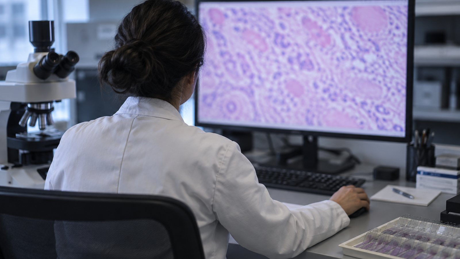
Once hospitals began scanning slides at high resolution, AI had something it could read. The files are enormous, and a suspicious cluster may occupy a tiny part of one. A tool that marks where a human should look twice could help with that search.
PathAI and Paige.AI are among the companies that have built AI systems for analyzing pathology slides. Paige received FDA clearance in 2021 for its prostate cancer detection tool, the first AI system cleared for primary diagnosis in pathology. Studies published in Nature Medicine showed that pathologists working with AI assistance caught more cancers than those working without it, particularly in subtle cases that a single reviewer might miss.
The AI systems highlight regions of interest on a slide for the pathologist to review. For someone reviewing fifty slides in a day, that triage can reduce the chance that a small cluster of abnormal cells gets overlooked.
The computer can also make the same mistake consistently. A threshold trained on different scanners or patient groups may stop working, so I would require local testing before deployment.
I would use it as a highlighter. Let the model point to the region it finds suspicious. Then let the pathologist decide what the cells mean in the rest of the case. That narrower job sounds much more useful than an “AI diagnoses cancer” headline.
The slide is only part of the case
A pathologist may compare several stains, the biopsy location and the patient's history before naming a disease. A model trained on one image type does not automatically understand the rest of that evidence.
There is also a practical question: what does the software do when the slide is folded, blurry or stained differently? A confident prediction on a bad image is worse than a system that admits it cannot read the case.
The hospital should track disagreements between the tool and its pathologists. Those cases are where hidden weaknesses appear. They are also where the software may catch a detail that deserves another look.